Extended Abstract
1. Introduction
Non-alcoholic fatty liver disease is a chronic liver disorder [1]. This metabolic disorder can affect mitochondrial dynamics [3]. The decrease in fusion proteins in fatty liver disease is due to an increase in fat, which disrupts mitochondrial function, loss of membrane potential, decreased oxygen, and increased ROS production [5, 6].
According to the various research, it has been reported that regular aerobic activity can reduce fat and liver vulnerability and reduce inflammation in non-alcoholic fatty liver. It has also been reported that resveratrol due to its polyphenol compounds can reduce the vulnerability of fatty liver and mitochondrial dynamics in cardiac myocytes [13].
The aim of this study was to investigate the effect of aerobic exercise supplemented with resveratrol on the expression of MFn1 and MFn2 in cardiac mitochondrial myocytes in a non-alcoholic fatty liver animal model.
2. Materials and Methods
In this experimental study, 48 Wistar eight-week-old male rats were selected as the sample and randomly divided into diseased group (NAFLD) and Control-healthy group (CN). Control rats were fed a standard diet; they were fed a high-fat diet for 6 weeks to induce NAFLD [21].
The diseased group rats itself were divided into 5 experimental subgroups: Diseased (NAFLD), Sham (SHAM), Practice-Patient (TRNAF), Supplement-Patient (SUPNAF), and Practice-Supplement-Patient (TRSUPNAF). The training program was eight weeks, starting at 15 m/min for the first week, 5 min for each session, and 1-2 m/min for each session. The time was also increased by 2-4 min, reaching at speed of 20 m/min in the fourth week; the time met 60 min and remained unchanged until the last week [22].
In the SUPNAF and TRSUPNAF groups, resveratrol was injected intraperitoneally at a dose of 20 mg/kg [23]. Real time PCR was used to evaluate the expression of MFn1 and MFn2. One-way ANOVA and Tukey post hoc test were used for statistical analysis at the significant level of P≤0.05.
3. Results
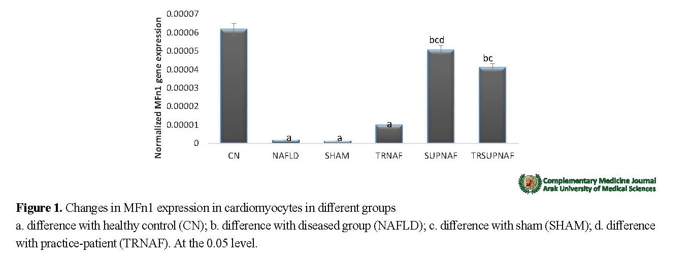
Data analysis showed that there was a significant difference in the rate of MFn1 expression of cardiomyocytes between different groups (F=11.743, P=0.000). The Tukey post hoc test showed significant differences between following groups: CN groups with NAFLD (P=0.001), SHAM (P=0.001), and TRNAF (P=0.001); between NAFLD group with SUPNAF (P=0.002) and TRSUPNAF (P=0.017); between SHAM group with SUPNAF (P=0.002) and TRSUPNAF (P=0.015); and also between TRNAF and TRSUPNAF group (P=0.013) (Figure 1).
Other results of this study showed a significant difference in the rate of Mfn2 expression of cardiomyocytes between different groups (P=0.000, F=27.482). The Tukey post hoc test showed significant differences between following groups: CN groups with NAFLD (P=0.001), SHAM (P=0.001) and TRNAF (P=0.001); between NAFLD group with SUPNAF (P=0.000) and TRSUPNAF (P=0.000); between SHAM group with SUPNAF (P=0.000) and TRSUPNAF (P=0.000); and also between TRNAF group with SUPNAF (0.000) and TRSUPNAF (P=0.000) (Figure 2).
4. Discussion
The results of this study showed that MFn1 and MFn2 expression was significantly decreased in the fatty liver mice compared to the healthy group. The reason for the decrease in these parameters may be due to the fusion protein imbalance. The decrease in the balance between fusion proteins in fatty liver disease is due to an increase in fat, which disrupts mitochondrial function [5, 6]. Excessive ROS induced by fatty liver can induce apoptosis and cardiac necrosis [26].
Liu et al. (2014) in a study on male fatty liver model rats reported a decrease in MFn1 and MFn2 [16]. In NAFLD, mitochondrial dysfunction inhibits MFn2, resulting in decreased mitochondrial respiration of cardiac myocytes and increased susceptibility to oxidative stress [11]. Other results of this study showed a significant increase in MFn1 and MFn2 expression in extract and exercise-extract groups compared to the diseased group.
Regular physical activity reduces inflammatory markers and increases capacity or antioxidant activity and reduces oxidative stress [5]. The decrease in oxidative stress indices is associated with increased expression of MFn1 and MFn2 genes, which enhances mitochondrial fusion activity [39].
Our findings indicate that administration of resveratrol along with aerobic exercise has a greater therapeutic effect on increased cardiac MFn1 and MFn2 expression than exercise alone. The use of resveratrol is associated with decreased ROS production and decreased expression of inflammatory cytokines as well as increased anti-inflammatory markers and improved antioxidant status [13]. Resveratrol appears to induce mitochondrial biogenesis by activating AMPK and mitochondrial transcription factors [43]. Aerobic exercise along with antioxidant supplements, including resveratrol, greatly help to inhibit the production of free radicals and oxidative stress in cardiac tissue cells of NAFLD patients, thereby enhancing the expression of fusion genes.

Ethical Considerations
Compliance with ethical guidelines
This study was conducted in accordance with the ethical standards of working with animals in accordance with the Ethics Committee of Islamic Azad University of Sari Branch: Code R.IAU.SARI.REC.1397.8.
Funding
The present paper was extracted from the PhD thesis of the first author, Department of Sport Physiology, Faculty of Physical Education and Sport Sciences Ayatollah Amoli Branch, Islamic Azad University, Amol.
Authors' contributions
Conceptualization, Methodology and Validation: Ahmad Abdi, Hasan Delroz, Parvin Farzanegi; Investigation, Resource, Original Draft Preparation: Ahmad Abdi, Hasan Delroz, Parvin Farzanegi, Alireza Barari; Editing and Review: Ahmad Abdi, Hasan Delroz; Supervision and Project Administration: Ahmad Abdi, Alireza Barari, Parvin Farzanegi, Funding Acquisition: Hasan Delroz.
Conflicts of interest
The authors declared no conflict of interest.
Acknowledgements
The authors would like to thank Islamic Azad University of Amol.