Write your message
Volume 10, Issue 4 (3-2021)                   cmja 2021, 10(4): 396-409 | Back to browse issues page


XML Persian Abstract Print


Download citation:
BibTeX | RIS | EndNote | Medlars | ProCite | Reference Manager | RefWorks
Send citation to:

Shirkhani Y, Peeri M, Azarbayjani M A, Matinhomaee H. Effect of Resistance Exercise and Vitamin C Intake on Expression of Telomerase Reverse Transcriptase and Telomere Repeat Binding Factor-2 Genes and the Diameter and Number of Myofibrils in Old Rats. cmja 2021; 10 (4) :396-409
URL: http://cmja.arakmu.ac.ir/article-1-789-en.html
1- Department of Sports Physiology, Faculty of Physical Education and Sports Sciences, Central Tehran Branch, Islamic Azad University, Tehran, Iran.
2- Department of Sports Physiology, Faculty of Physical Education and Sports Sciences, Central Tehran Branch, Islamic Azad University, Tehran, Iran. , m.peeri@gmail.com
Full-Text [PDF 5667 kb]   (1258 Downloads)     |   Abstract (HTML)  (2931 Views)
Full-Text:   (2886 Views)

1. Introduction

he aging cell has certain characteristics, the most important of which are increased cell size, distinct morphology, extensive changes in gene expression, and shortened telomere length [1]. Telomerase Reverse Transcriptase (TERT) is a catalytic subunit of enzyme telomerase. TERT is a protein with catalytic and transcriptase activity [2]. Other important proteins that play an important role in telomeric structure are telomere repeat binding factors (TRFs) with isoforms 1 and 2 and bind directly to TTAGGG repeats [3]. Oxidative pressure appears to affect the length of telomerase and myofibrils. 
Vitamin C is mixed with antioxidant activity. However, there are no clear results about the effect of vitamin C consumption on telomerase length as well as TERT and TRF2. Some studies have shown that long-term physical activity plays an important role in controlling aging by affecting chromosomes [4]. There are no clear results about the effect of resistance exercise as well as the effect of vitamin C intake and their combined effect on telomerase length and especially on TRF2 and TERT gene expression. In this regard, this study aims to evaluate the effect of resistance exercise along with liposomal vitamin C intake on the expression of TERT and TRF2 genes and on the diameter and number of myofibrils in old male Wistar rats.

2. Materials and Methods

This is an experimental study on 25 male Wistar rats aged 24 weeks and weighted 280-320 g. They were randomly divided into five groups of young control (n=5), old+resistance exercise (n=5), old+vitamin intake (n=5), old+resistance exercise+vitamin intake (n=5), and old control (n=5). In the exercise groups, 20 minutes of resistance exercise with ladders and weights were performed for 8 weeks, 3 days per week. At the end of the exercise period, rats in each group were examined for further studies. In the supplementation groups, liposomal vitamin C was administered daily by gavage per kg body weight. Shapiro-Wilk test was used to investigate the normality of data distribution. One-way ANOVA test was used to examine the differences between groups and Tukey’s post hoc test was used to determine where these differences were found. All analyses were performed in SPSS V. 22 software considering a significance level of 0.05.

3. Results

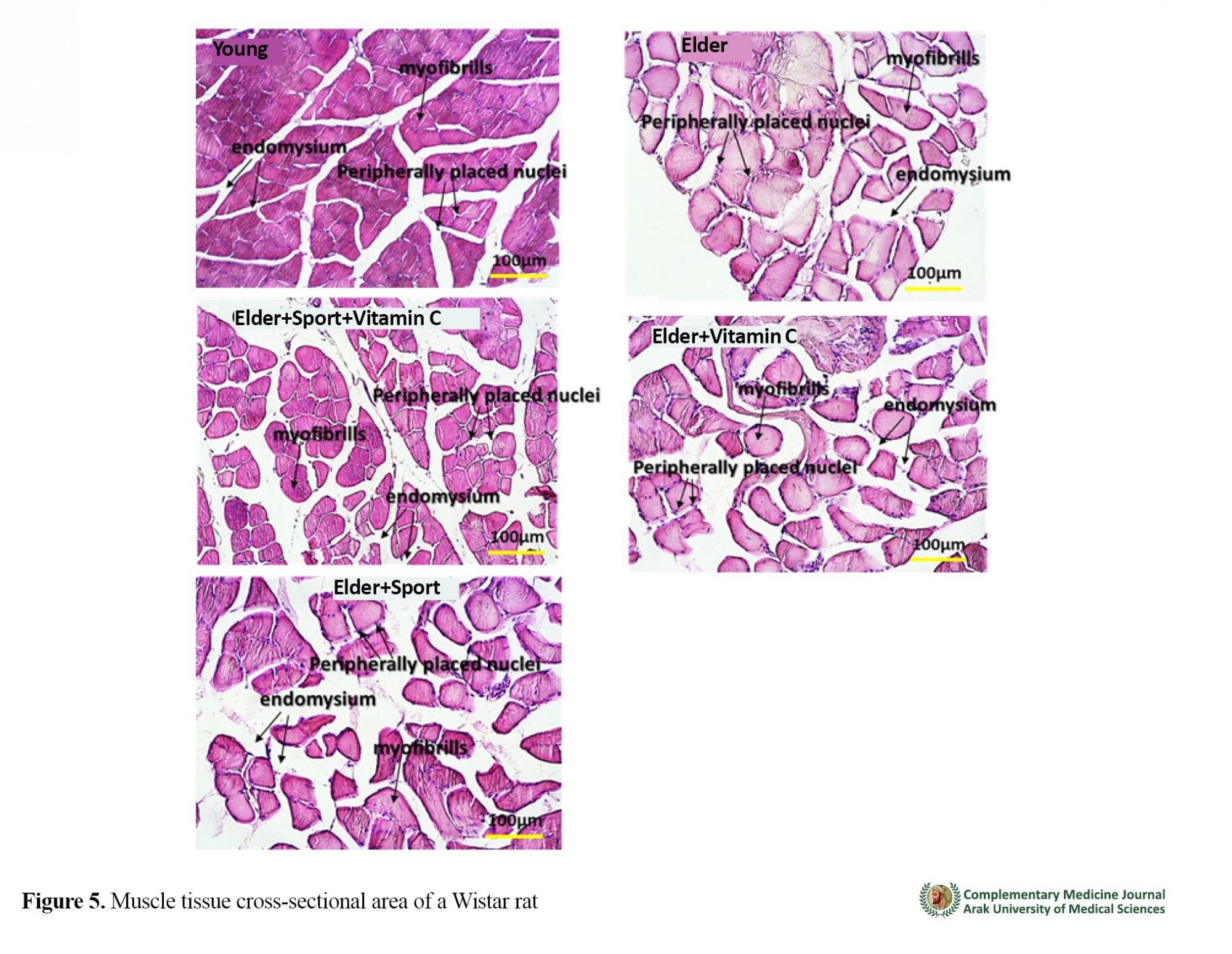
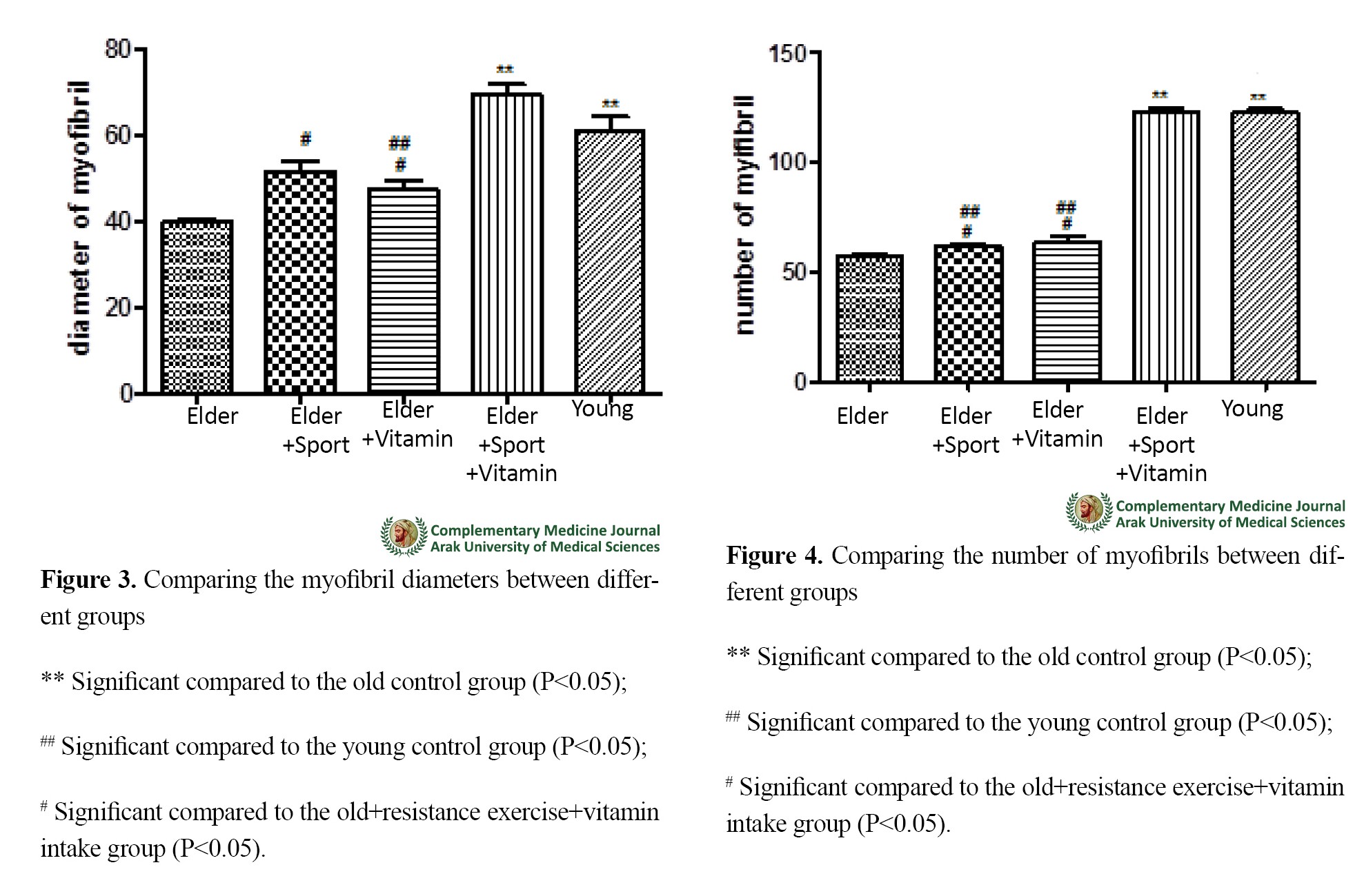
The results of ANOVA showed a significant difference between the groups in terms of TERT expression (P=0.001). The results of Tukey’s post hoc test showed that the significant differences were between the old control and young control groups (P=0.001). For TRF2 expression, ANOVA test results also showed a significant difference between the groups (P=0.001). Tukey’s post hoc test showed that the significant differences were between the old control and young control groups (P=0.001) and between the old+resistance exercise and young control groups (P=0.001). There was also a significant difference between the groups in terms of myofibril diameter (P=0.001). The results of Tukey’s post hoc test showed that the significant difference was between the young control and the old control groups, between the old+resistance exercise+vitamin intake and the old control groups (P=0.001), between the old+resistance exercise and the old+resistance exercise+vitamin intake groups (P=0.001), between the old+vitamin intake and the old+resistance exercise+vitamin intake groups, and between the old+vitamin intake and the young control groups (P=0.001). There was also a significant difference between the groups in terms of the number of myofibrils (P=0.001). The results of Tukey’s post hoc test showed that the significant difference was between the young control and the old control groups, between the old+resistance exercise+vitamin intake and the old control groups (P=0.001), between the old+resistance exercise and the old+resistance exercise+vitamin intake groups (P=0.001), between the old+resistance exercise and the young control groups (P=0.001), between the old+vitamin intake and the old+resistance exercise+vitamin intake groups, and between the old+vitamin intake and the young control groups (P=0.001) (Figures 1, 2, 3, 4 & 5).


4. Conclusion

The purpose of this study was to investigate the effect of resistance exercise along with vitamin C consumption on the expression of TERT, TRF2 genes and the diameter and number of skeletal muscle myofibrils in old male Wistar rats. The results showed that there was a significant difference between the old and young control groups. However, no significant difference was observed between the other groups. Regarding TRF2 level, a significant difference was observed between the old and young control group (P=0.001) and between the trained old group and the young control group. in a study, rats with Manganese superoxide dismutase deficiency in muscle or heart tissue had inhibited telomerase activity and decreased expression of TERT and TRF2. However, antioxidant supplements prevented this process [5]. Part of the adaptation of telomerase to exercise should be attributed to the MAPK pathways. Activation of MAPK leads to a change in the positional activity of a variety of transcription factors that are associated with altered expression of telomere gene components [6]. On the other hand, exercise increases the antioxidant content and reduces the oxidative pressure [7]. It seems that the interactive effect of exercise and vitamin C intake is effective in reducing the maximum oxidative pressure on telomere length.
In the present study, the expression of TERT and TRF2, and the diameter and number of myofibrils in the group of old rats that did resistance exercise and took vitamin C, was close to those of young control rats. Increased TERT and especially TRF2 levels can be considered as the reasons for the increase in diameter and number of myofibrils in old rats. Due to the limited research on TERT and TRF2 levels in the elderly, as well as the effects of vitamin C and resistance exercise on it, no other rational mechanisms can be mentioned.
Eight weeks of resistance exercise along with vitamin C supplementation can significantly increase the number and diameter of skeletal muscle myofibrils in older rats.

Ethical Considerations

Compliance with ethical guidelines

This study was approved by the Ethics Committee of Sport Sciences Research Institute of Iran (Code: IR.SSRI.REC.1399.115). 

Funding

This study was extracted from a PhD. dissertation of the first author, at the Department of Sports Physiology, Faculty of Physical Education and Sports Sciences, Islamic Azad University, Central Tehran Branch, Tehran.

Authors' contributions

Performing training, writing, editing & review: Yousef shirkhani; Data analysis: Mohammad ali Azarbayjani; Editing & review: Hassan Matinhomaee, Maghsoud Peeri.

Conflicts of interest

The authors declared no conflict of interest.
Type of Study: Research | Subject: Other cases

References
1. Miljkovic N, Lim J-Y, Miljkovic I, Frontera WR. Aging of skeletal muscle fibers. Annals of rehabilitation medicine. 2015;39(2):155. [DOI:10.5535/arm.2015.39.2.155] [PMID] [PMCID]
2. Morsiani C, Bacalini MG, Santoro A, Garagnani P, Collura S, D'Errico A, et al. The peculiar aging of human liver: A geroscience perspective within transplant context. Ageing research reviews. 2019;51:24-34. [DOI:10.1016/j.arr.2019.02.002] [PMID]
3. Ho RT, Chan JS, Wang C-W, Lau BW, So KF, Yuen LP, et al. A randomized controlled trial of qigong exercise on fatigue symptoms, functioning, and telomerase activity in persons with chronic fatigue or chronic fatigue syndrome. Annals of Behavioral Medicine. 2012;44(2):160-70. [DOI:10.1007/s12160-012-9381-6] [PMID] [PMCID]
4. Pejenaute Á, Cortés A, Marqués J, Montero L, Beloqui Ó, Fortuño A, et al. NADPH oxidase overactivity underlies telomere shortening in human atherosclerosis. International Journal of Molecular Sciences. 2020;21(4):1434. [DOI:10.3390/ijms21041434] [PMID] [PMCID]
5. Heidenreich B, Kumar R. TERT promoter mutations in telomere biology. Mutation Research/Reviews in Mutation Research. 2017;771:15-31. [DOI:10.1016/j.mrrev.2016.11.002] [PMID]
6. Slattery ML, Herrick JS, Pellatt AJ, Wolff RK, Mullany LE. Telomere length, TERT, and miRNA expression. PLoS One. 2016;11(9):e0162077. [DOI:10.1371/journal.pone.0162077] [PMID] [PMCID]
7. Monaghan P, Eisenberg DT, Harrington L, Nussey D. Understanding diversity in telomere dynamics. The Royal Society; 2018. [DOI:10.1098/rstb.2016.0435] [PMID] [PMCID]
8. Prasad KN, Wu M, Bondy SC. Telomere shortening during aging: Attenuation by antioxidants and anti-inflammatory agents. Mechanisms of ageing and development. 2017;164:61-6. [DOI:10.1016/j.mad.2017.04.004] [PMID]
9. de Vos-Houben JM, Ottenheim NR, Kafatos A, Buijsse B, Hageman GJ, Kromhout D, et al. Telomere length, oxidative stress, and antioxidant status in elderly men in Zutphen and Crete. Mechanisms of ageing and development. 2012;133(6):373-7. [DOI:10.1016/j.mad.2012.04.003] [PMID]
10. Kiecolt-Glaser JK, Epel ES, Belury MA, Andridge R, Lin J, Glaser R, et al. Omega-3 fatty acids, oxidative stress, and leukocyte telomere length: a randomized controlled trial. Brain, behavior, and immunity. 2013;28:16-24. [DOI:10.1016/j.bbi.2012.09.004] [PMID] [PMCID]
11. Bruns DR, Ehrlicher SE, Khademi S, Biela LM, Peelor III FF, Miller BF, et al. Differential effects of vitamin C or protandim on skeletal muscle adaptation to exercise. Journal of Applied Physiology. 2018;125(2):661-71. [DOI:10.1152/japplphysiol.00277.2018] [PMID] [PMCID]
12. Negaresh R, Ranjbar R, Habibi A, Gharibvand MM. The relationship between muscle volume and strength and some factors associated with sarcopenia in old men compared with young men. Zanko Journal of Medical Sciences. 2016;17(54):23-34.
13. Harris SE, Deary IJ, MacIntyre A, Lamb KJ, Radhakrishnan K, Starr JM, et al. The association between telomere length, physical health, cognitive ageing, and mortality in non-demented older people. Neuroscience letters. 2006;406(3):260-4. [DOI:10.1016/j.neulet.2006.07.055] [PMID]
14. Jiang H, Schiffer E, Song Z, Wang J, Zürbig P, Thedieck K, et al. Proteins induced by telomere dysfunction and DNA damage represent biomarkers of human aging and disease. Proceedings of the National Academy of Sciences. 2008;105(32):11299-304. [DOI:10.1073/pnas.0801457105] [PMID] [PMCID]
15. Negaresh R, Ranjbar R, Habibi A, Gharibvand MM. The effects of eight weeks of resistance training on some muscle hypertrophy and physiological parameters in elderly men. Journal of Geriatric Nursing. 2016;3(1):62-75. [DOI:10.21859/jgn.3.1.62]
16. Rae DE, Vignaud A, Butler-Browne GS, Thornell L-E, Sinclair-Smith C, Derman EW, et al. Skeletal muscle telomere length in healthy, experienced, endurance runners. European journal of applied physiology. 2010;109(2):323-30. [DOI:10.1007/s00421-010-1353-6] [PMID]
17. Ponsot E, Lexell J, Kadi F. Skeletal muscle telomere length is not impaired in healthy physically active old women and men. Muscle & nerve. 2008;37(4):467-72. [DOI:10.1002/mus.20964] [PMID]
18. Werner C, Fürster T, Widmann T, Pöss J, Roggia C, Hanhoun M, et al. CLINICAL PERSPECTIVE. Circulation. 2009;120(24):2438-47. [DOI:10.1161/CIRCULATIONAHA.109.861005] [PMID]
19. Ludlow AT, Witkowski S, Marshall MR, Wang J, Lima LC, Guth LM, et al. Chronic exercise modifies age-related telomere dynamics in a tissue-specific fashion. Journals of Gerontology Series A: Biomedical Sciences and Medical Sciences. 2012;67(9):911-26. [DOI:10.1093/gerona/gls002] [PMID] [PMCID]
20. Eskandari A, Fashi M, Dakhili AB. Effect of high intensity interval and continuous endurance training on TRF2 and TERT gene expression in heart tissue of aging male rats. Journal of Gorgan University of Medical Sciences. 2019;21(2).
21. Thirupathi A, da Silva Pieri BL, Queiroz JAMP, Rodrigues MS, de Bem Silveira G, de Souza DR, et al. Strength training and aerobic exercise alter mitochondrial parameters in brown adipose tissue and equally reduce body adiposity in aged rats. Journal of physiology and biochemistry. 2019;75(1):101-8. [DOI:10.1007/s13105-019-00663-x] [PMID]
22. Scheffer DL, Silva LA, Tromm CB, da Rosa GL, Silveira PC, de Souza CT, et al. Impact of different resistance training protocols on muscular oxidative stress parameters. Applied Physiology, Nutrition, and Metabolism. 2012;37(6):1239-46. [DOI:10.1139/h2012-115] [PMID]
23. Damas F, Phillips SM, Libardi CA, Vechin FC, Lixandrão ME, Jannig PR, et al. Resistance training‐induced changes in integrated myofibrillar protein synthesis are related to hypertrophy only after attenuation of muscle damage. The Journal of physiology. 2016;594(18):5209-22. [DOI:10.1113/JP272472] [PMID] [PMCID]
24. Gabrial SG, Shakib M-CR, Gabrial GN. Protective Role of Vitamin C Intake on Muscle Damage in Male Adolescents Performing Strenuous Physical Activity. Open access Macedonian journal of medical sciences. 2018(6(9:1594. [DOI:10.3889/oamjms.2018.337] [PMID] [PMCID]
25. Haendeler J, Hoffmann Jr, Diehl JF, Vasa M, Spyridopoulos I, Zeiher AM, et al. Antioxidants inhibit nuclear export of telomerase reverse transcriptase and delay replicative senescence of endothelial cells. Circulation research. 2004;94(6):768-75. [DOI:10.1161/01.RES.0000121104.05977.F3] [PMID]
26. Makino N, Maeda T, Oyama J-i, Sasaki M, Higuchi Y, Mimori K, et al. Antioxidant therapy attenuates myocardial telomerase activity reduction in superoxide dismutase-deficient mice. Journal of molecular and cellular cardiology. 2011;50(4):670-7. [DOI:10.1016/j.yjmcc.2010.12.014] [PMID]
27. Rose BA, Force T, Wang Y. Mitogen-activated protein kinase signaling in the heart: angels versus demons in a heart-breaking tale. Physiological reviews. 2010;90(4):1507-46. [DOI:10.1152/physrev.00054.2009] [PMID] [PMCID]
28. Baghaiee B, Karimi P, Siahkouhian M, Pescatello LS. Moderate aerobic exercise training decreases middle-aged induced pathologic cardiac hypertrophy by improving Klotho expression, MAPK signaling pathway and oxidative stress status in Wistar rats. Iranian Journal of Basic Medical Sciences. 2018;21(9):911-9.

Add your comments about this article : Your username or Email:
CAPTCHA

Send email to the article author


Rights and permissions
Creative Commons License This work is licensed under a Creative Commons Attribution-NonCommercial 4.0 International License.

© 2025 CC BY-NC 4.0 | Complementary Medicine Journal

Designed & Developed by : Yektaweb